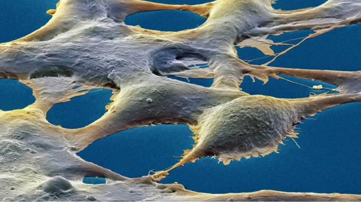
Kidney cells possess the ability to store memories, albeit in a metaphorical sense. Historically, neurons have been the primary cell type associated with memory. However, researchers have discovered that kidney cells, far removed from the brain, can also store information and recognize patterns in a manner similar to neurons. This finding was published on November 7 in Nature Communications.
“We’re not suggesting that this type of memory aids in learning trigonometry or remembering how to ride a bike, nor does it store childhood memories,” explains Nikolay Kukushkin, a neuroscientist at New York University. “This research expands our understanding of memory without challenging the existing conceptions of memory within the brain.”
In their experiments, kidney cells exhibited signs of what is known as the “massed-space effect.” This well-documented feature of memory in the brain involves storing information in small, manageable chunks over time rather than all at once. Cells, regardless of their type, need to keep track of various elements, and one way they do this is through a protein crucial for memory processing called CREB. This protein, along with other molecular components of memory, is found in both neurons and nonneuronal cells.
While both types of cells contain similar components, researchers were uncertain whether these components functioned in the same way. In neurons, when a chemical signal is received, the cell begins producing CREB. This protein then activates more genes that further modify the cell, initiating the molecular memory process. Kukushkin and his team aimed to determine if CREB in nonneuronal cells responds to incoming signals in a similar manner.
The researchers introduced an artificial gene into human embryonic kidney cells. This gene closely resembles the naturally occurring DNA sequence that CREB binds to, which the researchers refer to as a memory gene. The inserted gene also included instructions for producing a glowing protein found in fireflies. The team then observed how the cells responded to artificial chemical pulses that mimic the signals triggering memory machinery in neurons.
“Depending on the amount of light produced by the glowing protein, we can gauge how strongly the memory gene was activated,” Kukushkin explains. Different timing patterns of pulses led to varied responses. When the researchers applied four three-minute chemical pulses separated by 10 minutes, the light emitted 24 hours later was stronger than in cells subjected to a single 12-minute pulse.
“This massed-spaced effect has never been observed outside the brain; it has always been considered a property of neurons and the brain’s memory formation process,” Kukushkin notes. “However, we propose that if non-brain cells are given sufficiently complex tasks, they too may be able to form a memory.”
Neuroscientist Ashok Hegde finds the study intriguing, as it applies a neuroscience principle broadly to understand gene expression in nonneuronal cells. However, it remains unclear how widely these findings can be applied to other cell types, according to Hegde of Georgia College & State University in Milledgeville. Nonetheless, he believes this research could potentially aid in the search for drugs to treat human diseases, particularly those involving memory loss.
Kukushkin concurs, suggesting that the body’s ability to store information could have significant implications for health. “Perhaps we can consider cancer cells as having memories and think about what they might learn from the pattern of chemotherapy,” he says. “Maybe we need to consider not only the dosage of a drug but also its timing pattern, similar to how we think about learning more efficiently.”
Source link: https://www.sciencenews.org


