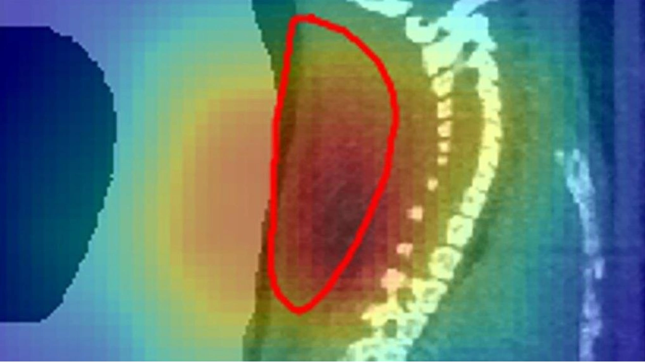
Particle beams, capable of delivering a concentrated burst of destructive energy directly to tumors, have been successfully tracked during treatment. This precision is crucial, especially when the tumor is located near sensitive organs like the spinal cord or brain stem. Researchers have now achieved the first successful treatment of tumors in mice using a radioactive beam, as detailed in a paper submitted on September 23 to arXiv.org.
The technique holds promise for future human treatments with millimeter precision. Unlike X-rays, which can cause collateral damage along their path, particle beams like protons or ions deposit most of their energy in a single spot. Ion treatment centers currently use stable, non-radioactive ions such as carbon-12. However, body movements and organ shifts can complicate targeting. Real-time confirmation of the beam's position is essential, and this new method provides just that.
Physicist Marco Durante of GSI Helmholtz Centre for Heavy Ion Research in Darmstadt, Germany, explains that using radioactive ions like carbon-11 allows simultaneous tumor destruction and beam visualization. Carbon-11 emits positrons upon decay, which can be detected via positron emission tomography (PET). In the study, carbon-11 ions were used to treat mice with spinal tumors, and the beam's position was accurately confirmed during treatment, leading to tumor shrinkage.
Previous attempts to use PET with stable ions were challenging due to the low number of radioactive fragments. Radioactive ion beams, however, emit many more positrons, providing a clear image of the beam's stopping point. This technique could also help scientists track how radioactive material moves through the body after treatment, potentially revealing if blood vessels are being damaged, thereby cutting off the tumor's energy supply.


